Beranda
/ Animal Cell Telophase Diagram : The Diagram Given Below Represents A Stage During Cell Division Identify Whether It Is A Plant Cell Or An Animal Cell Give A Reason In Support Of Your Answer And Name The : Cytoplasm, ribosomes, rough endoplasmic reticulum;
Animal Cell Telophase Diagram : The Diagram Given Below Represents A Stage During Cell Division Identify Whether It Is A Plant Cell Or An Animal Cell Give A Reason In Support Of Your Answer And Name The : Cytoplasm, ribosomes, rough endoplasmic reticulum;
Animal Cell Telophase Diagram : The Diagram Given Below Represents A Stage During Cell Division Identify Whether It Is A Plant Cell Or An Animal Cell Give A Reason In Support Of Your Answer And Name The : Cytoplasm, ribosomes, rough endoplasmic reticulum;. Lets us discuss the animal cell, types of an animal cell, animal cell diagram, its all cell organelles are marked clearly in the diagram. What invention made it possibe for people to study cells? During this phase, the nuclear membrane reforms, the nucleolus reappears, and the although the overall process of splitting is the same, it looks a bit different in animal cells and plant cells. Cell cycle lab slides use the following slides to identify the various phases of mitosis. Learners need to know the names of the phases and they need to be able to draw simple descriptive diagrams showing the chromosome changes.
Mitosis is nuclear division plus cytokinesis, and produces two identical daughter cells during prophase, prometaphase, metaphase, anaphase, and telophase. Read more about animal cell, functions and structure of animal. Animal cell diagram simple gcse. So that from one primary oocyte a single ovum is produced (see diagram of oogenesis immediately below). During this phase, the nuclear membrane reforms, the nucleolus reappears, and the although the overall process of splitting is the same, it looks a bit different in animal cells and plant cells.
Mitosis Biol110f2012 Confluence from wikispaces.psu.edu During telophase, the effects of prophase and prometaphase (the nucleolus and nuclear membrane disintegrating) are reversed. Lets us discuss the animal cell, types of an animal cell, animal cell diagram, its all cell organelles are marked clearly in the diagram. This furrow deepens and considerable movement of cytoplasm. The file size of this svg image may be irrationally large because its text has been converted to paths, to inhibit translation. Under the microscope, an animal cell shows many different parts called organelles, that work together to keep the cell functional. Mitosis is nuclear division plus cytokinesis, and produces two identical daughter cells during prophase, prometaphase, metaphase, anaphase, and telophase. Simplified diagram of an animal cell. Telophase is the final step of mitosis.
Some students may be able to identify some of the structures.
Polish your personal project or design with these telophase transparent png images, make it even more personalized and more attractive. The cell surface membrane pinches inwards creating a cleavage furrow in the middle of the cell which contracts, dividing the cytoplasm anaphase ii: Both plant and animal cells are surrounded by a cell membrane composed of lipids and proteins. Cell cycle lab slides use the following slides to identify the various phases of mitosis. All cells are produced from other cells. Let us look at animal cell parts and functions, using diagrams and illustrations. Lets us discuss the animal cell, types of an animal cell, animal cell diagram, its all cell organelles are marked clearly in the diagram. Telophase (events of prophase reversed) a. Although animal cells lack these cell structures, both of them have nucleus, mitochondria, endoplasmic reticulum, etc. A comparison of plant and animal cells using labelled diagrams and descriptive explanations. It helps in carrying out the functions such as respiration, nutrition, digestion, excretion etc. The cell cycle and cell division. Under the microscope, an animal cell shows many different parts called organelles, that work together to keep the cell functional.
Both plant and animal cells are surrounded by a cell membrane composed of lipids and proteins. That cells can be of different shapes and sizes. During telophase ii, the sister chromatids reach opposite poles, cytokinesis occurs, the two cells produced by meiosis i divide to form four haploid daughter cells, and nuclear envelopes form. Here you can explore hq telophase transparent illustrations, icons and clipart with filter setting like size, type, color etc. Under the microscope, an animal cell shows many different parts called organelles, that work together to keep the cell functional.
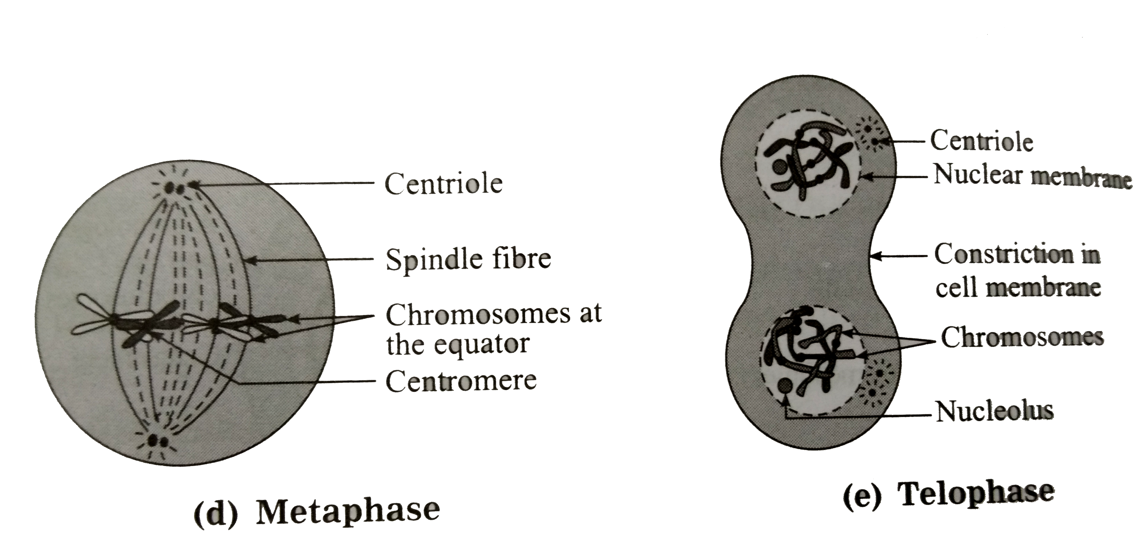
With The Help Of Suitasble Diagrams Explain In Mitosis In Detail from d10lpgp6xz60nq.cloudfront.net The chromosomes form a loose during the end of telophase a furrow is formed in the cell membrane along the equator. Nuclei are forming around the 4 groups of condensed. The cell surface membrane pinches inwards creating a cleavage furrow in the middle of the cell which contracts, dividing the cytoplasm anaphase ii: The cell cycle and cell division. The animal cell diagram on the free worksheet will teach students to identify the function of the major parts of the animal cell. After completing this section, you should know: Cytoplasm, ribosomes, rough endoplasmic reticulum; From wikimedia commons, the free media repository.
Simplified diagram of an animal cell.
Telophase is the final step of mitosis. Polish your personal project or design with these telophase transparent png images, make it even more personalized and more attractive. Venn diagram comparing plant cells and animal file size: Cytoplasm, ribosomes, rough endoplasmic reticulum; After completing this section, you should know: All cells are produced from other cells. An animal cell is the smallest unit that makes up the varied tissues of animal species. Nuclear membrane and nucleolus reappear. During telophase ii, the sister chromatids reach opposite poles, cytokinesis occurs, the two cells produced by meiosis i divide to form four haploid daughter cells, and nuclear envelopes form. It's a completely free picture material come from the public internet and the real upload of users. Although animal cells lack these cell structures, both of them have nucleus, mitochondria, endoplasmic reticulum, etc. This animal cell diagram doesn't represent any particular animal cell, it provides insight into the primary. This furrow deepens and considerable movement of cytoplasm.
Some students may be able to identify some of the structures. Both plant and animal cells are surrounded by a cell membrane composed of lipids and proteins. Plant cell and animal cell fall under eukaryotic type. Telophase is the final stage in both meiosis and mitosis in a eukaryotic cell. Have cell walls and chloroplasts in contrast to animal cells which have no cell wall or chloroplasts.
Draw A Neat Labeled Diagram To Show The Metaphase Stage Of Mitosis In An Animal Cell Having 6 Chromosome Studyrankersonline from i1.wp.com There are hundreds of cell types in a developed organism, which are specific to their location and function. Venn diagram comparing plant cells and animal file size: All cells are produced from other cells. It's a completely free picture material come from the public internet and the real upload of users. Simplified diagram of an animal cell. Plant cell and animal cell fall under eukaryotic type. The resolution of image is 522x385 and classified to mouse animal, animal crossing, animal. Nuclei are forming around the 4 groups of condensed.
Here you can explore hq telophase transparent illustrations, icons and clipart with filter setting like size, type, color etc.
The file size of this svg image may be irrationally large because its text has been converted to paths, to inhibit translation. The resolution of image is 522x385 and classified to mouse animal, animal crossing, animal. Venn diagram comparing plant cells and animal file size: Animal cell diagram simple gcse. Some students may be able to identify some of the structures. Cytoplasm, ribosomes, rough endoplasmic reticulum; The red blood cells make up the blood, while the nerve cells make up the nervous system tissues. The correct diagram of animal cell is. Telophase is the final step of mitosis. So that from one primary oocyte a single ovum is produced (see diagram of oogenesis immediately below). The role and function of the plasma membrane; Cells are the basic units of structure and function in living things. Both plant and animal cells are surrounded by a cell membrane composed of lipids and proteins.
Berbagi :
Posting Komentar
untuk "Animal Cell Telophase Diagram : The Diagram Given Below Represents A Stage During Cell Division Identify Whether It Is A Plant Cell Or An Animal Cell Give A Reason In Support Of Your Answer And Name The : Cytoplasm, ribosomes, rough endoplasmic reticulum;"


Posting Komentar untuk "Animal Cell Telophase Diagram : The Diagram Given Below Represents A Stage During Cell Division Identify Whether It Is A Plant Cell Or An Animal Cell Give A Reason In Support Of Your Answer And Name The : Cytoplasm, ribosomes, rough endoplasmic reticulum;"